
Med Oral Patol Oral Cir Bucal. 2015 Nov 1;20 (6):e685-92. Oral leukoplakia
e685
Journal section: Oral Medicine and Pathology
Publication Types: Review
Oral leukoplakia, the ongoing discussion on denition and terminology
Isaäc van der Waal
VU University Medical Center (VUmc)/Academic Centre for Dentistry Amsterdam (ACTA), Department of Oral and Maxil-
lofacial Surgery and Oral Pathology, P.O. Box 7057, 1007 MB Amsterdam, The Netherlands
Correspondence:
Department of Oral and Maxillofacial Surgery and Oral Pathology
VU University Medical Center
P.O. Box 7057
1007 MB Amsterdam
The Netherlands
I.vanderwaal@vumc.nl
Received: 17/08/2015
Accepted: 27/09/2015
Abstract
In the past decades several denitions of oral leukoplakia have been proposed, the last one, being authorized by
the World Health Organization (WHO), dating from 2005. In the present treatise an adjustment of that denition
and the 1978 WHO denition is suggested, being : “A predominantly white patch or plaque that cannot be char-
acterized clinically or pathologically as any other disorder; oral leukoplakia carries an increased risk of cancer
development either in or close to the area of the leukoplakia or elsewhere in the oral cavity or the head-and-neck
region”. Furthermore, the use of strict diagnostic criteria is recommended for predominantly white lesions for
which a causative factor has been identied, e.g. smokers’ lesion, frictional lesion and dental restoration associat-
ed lesion. A nal diagnosis of such leukoplakic lesions can only be made in retrospect after successful elimination
of the causative factor within a somewhat arbitrarily chosen period of 4-8 weeks. It seems questionable to exclude
“frictional keratosis” and “alveolar ridge keratosis” from the category of leukoplakia as has been suggested in
the literature. Finally, brief attention has been paid to some histopathological issues that may cause confusion in
establishing a nal diagnosis of leukoplakia.
Key words: Oral leukoplakia, potentially malignant oral disorders, denition.
van der Waal I. Oral leukoplakia, the ongoing discussion on denition and
terminology. Med Oral Patol Oral Cir Bucal. 2015 Nov 1;20 (6):e685-92.
http://www.medicinaoral.com/medoralfree01/v20i6/medoralv20i6p685.pdf
Article Number: 21007 http://www.medicinaoral.com/
© Medicina Oral S. L. C.I.F. B 96689336 - pISSN 1698-4447 - eISSN: 1698-6946
eMail: [email protected]
Indexed in:
Science Citation Index Expanded
Journal Citation Reports
Index Medicus, MEDLINE, PubMed
Scopus, Embase and Emcare
Indice Médico Español
doi:10.4317/medoral.21007
http://dx.doi.org/doi:10.4317/medoral.21007
Introduction
1. The denition and terminology of oral leukoplakia
and leukoplakialike (“leukoplakic”) lesions and dis-
orders of the oral mucosa is the subject of discussion
in the literature for many decades. This discussion is
mainly focused on clinical aspects, but is partly related
to some histopathological aspects as well. In this trea-
tise the various denitions of oral leukoplakia will be
discussed, resulting in a suggestion for a slight adjust-
ment of the 2005 WHO denition. Furthermore, some
leukoplakic lesions will be discussed that may cause
some confusion as whether or not to exclude them from
the category of leukoplakia; examples are “alveolar
ridge keratosis” and “frictional keratosis”.
Med Oral Patol Oral Cir Bucal. 2015 Nov 1;20 (6):e685-92. Oral leukoplakia
e686
Denition
2.1 The various WHO denitions and suggestion for an
adjusted denition
In 1978, oral leukoplakia has been dened by the World
Health Organization (WHO) as: ‘A white patch or plaque
that cannot be characterized clinically or pathologically
as any other disease’ (1). In an explanatory note it has
been explicitly stated that the term leukoplakia is unre-
lated to the absence or presence of epithelial dysplasia.
In a monograph by the WHO, published in 1997, the
phrase: ‘any other denable disease’ was replaced by
‘any other denable lesion’ (2). No justication has been
provided for this change.
In 2002, it has been recommended to make a distinction
between a provisional clinical diagnosis of oral leuko-
plakia and a denitive one (Table 1) (3). A provisional
diagnosis was made when a lesion at the initial clinical
examination could not be clearly diagnosed as either leu-
koplakia or any other disease. In case of a provisional
clinical diagnosis, Certainty factor 1 was assigned (Ta-
ble 2). A denitive clinical diagnosis of leukoplakia was
made after unsuccessful elimination of suspected etio-
logical factors or in the absence of such factors, assigning
Certainty factor 2. Certainty factor 3 was assigned when
histopathological examination of an incisional biopsy did
not show the presence of any other diseases. In case of an
excisional biopsy or surgical excision, performed after an
incisional biopsy, Certainty factor 4 was assigned based
on histopathological examination of the surgical speci-
men (Fig. 1). It goes without saying that in epidemiologi-
cal studies a Certainty factor 1, based on a single oral
examination, is acceptable, while in scientic studies,
e.g. comparing different treatment results, Certainty fac-
tor 4 will be required, if feasible. Apparently, the recom-
mendation to use a Certainty factor has not been widely
accepted in the recent literature (4), although the use of
such factor is common practice in cancer registries.
In 2005, the denition of oral leukoplakia has been
changed at a WHO supported meeting into: ‘A white
plaque of questionable risk having excluded (other)
known diseases or disorders that carry no increased risk
for cancer’ (5). During the latter meeting it has deliber-
ately been decided to consider leukoplakia a potentially
malignant- premalignant and precancerous are equiva-
lent adjectives- disease and not a lesion since it is well
known that cancer development not always occurs in or
close to the leukoplakia but may also occur at other sites
in the oral cavity or the head-and-neck region.
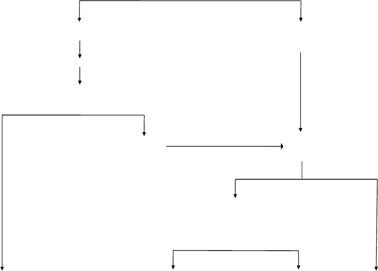
DIAGNOSIS OF ORAL LEUKOPLAKIA
(Provisional clinical diagnosis, C 1*)
Consider the taking of a biopsy, particularly in case of symptoms No possible cause(s)
(Definitive clinical diagnosis, C 2)
In the absence of dysplasia
Elimination of possible cause(s), such as mechanical irritation, amalgam restoration
in direct contact with the white lesion, fungal infection, and tobacco habits
(maximum 4-8 weeks to observe the result)
No response Biopsy
(Definitive clinical diagnosis, C 2)
Good response Definitive clinicopathological diagnosis
C 3 (in case of incisional biopsy only
C 4 (in case of excisional biopsy or surgical
excision after an incisional biopsy
Definable lesion Non-dysplastic leukoplakia Dysplastic leukoplakia Definable lesion
* C=Certainty factor (see table 2)
Table 1. Diagnosis of oral leukoplakia.
Med Oral Patol Oral Cir Bucal. 2015 Nov 1;20 (6):e685-92. Oral leukoplakia
e687
Although various international meetings on the sub-
ject of oral leukoplakia have been held between 1978
(WHO) and 2005 (WHO) no substantial changes in the
denition of leukoplakia have resulted from these meet-
ings.
In the sixties of the past century a minimum size of 5
mm was required before being allowed to use the term
leukoplakia (6). There seems no strong reason to re-
introduce a minimum size, since cancer devopment
may also take place in very small leukoplakias. Another
part of previous denitions of leukoplakia has been the
requirement of a non-removable nature of the white le-
sion, apparently mainly meant to separate pseudomem-
branous candidiasis from leukoplakia. The adjectives
“non-removable” or “non-scrapable” seem, indeed, to
have some practical value, but there is no strong reason
to include these in the denition.
The advantage of the 2005 WHO denition above the
one from 1978 is its statement about the behaviour of
oral leukoplakia (“questionable risk”). Unfortunately, in
both WHO denitions (1978 and 2005) the diagnosis of
leukoplakia is one by exclusion (of other “known dis-
eases or disorders”). A list of these “known diseases”
is depicted in table 3. Some of these diseases will be
briey commented upon (see ad 3). Many, if not most,
of the listed entities may be easy to distinguish from
leukoplakia by an experienced clinician, either based on
the history or based on the clinical appearance, but this
may not be the case for the less experienced clinician,
being either a dentist, an oral and maxillofacial surgeon,
an otolaryngologist or a dermatologist. Furthermore, it
does not seem realistic to expect family doctors to be
knowledgable in this eld, since probably in most parts
of the world little attention is paid to oral diseases dur-
ing the medical curriculum. One may even discuss at
what level dentists-general practitioners should be edu-
cated in the diagnosis and management of the numerous
lesions and disorders of the oral mucosa .
A combination of the 1978 and the 2005 WHO de-
nitions of oral leukoplakia may result in the following
text: “A predominantly white patch or plaque that can-
not be characterized clinically or pathologically as any
other disorder; oral leukoplakia carries an increased risk
of cancer development either in or close to the area of
leukoplakia or elsewhere in the oral cavity or the head-
and-neck region”.
2.2 What is a signicant increased risk of cancer de-
velopment?
In dening a potentially malignant disorder it is usu-
ally stated that there is “signicant increased risk” of
cancer development, without specifying “signicant”.
When the incidence- number of new patients per year-
of oral cancer is set at a low 2:100,000 population and
the annual malignant transformation rate of leukoplakia
at 2:100 (irrespective of the discussion whether or not
treatment of leukoplakia reduces the risk of malignant
transformation) there is a thousandfold risk in leukopla-
kia patients to develop cancer in comparison with pa-
tients not having leucoplakia. Probably, a thousandfold
increased risk is perceived, particularly by patients, as
being signicant.
Discussion on some “other known diseases and
disorders” that may have a leukoplakic appear-
ance
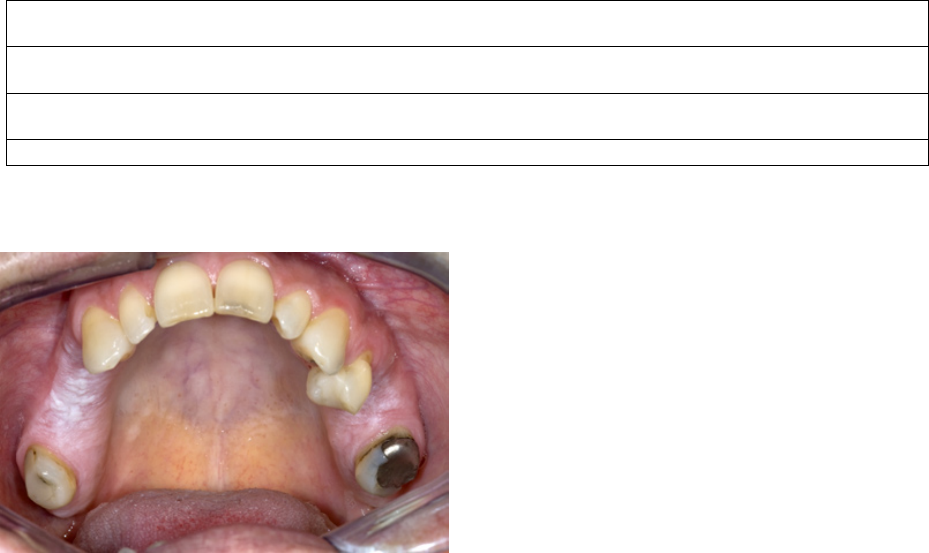
-3.1 Alveolar ridge “keratosis”
A few papers have been devoted to “alveolar ridge kera-
tosis” (7,8). Apparently, the supposed cause of the lesion
is chronic frictional (masticatory) trauma to the max-
1
C
1
Evidence from a single visit, applying inspection and palpation as the only diagnosis means (Provisional clinical
diagnosis), including a clinical picture of the lesion.
C
2
Evidence obtained by a negative result of elimination of suspected etiologic factors, e.g. mechanical irritation, during a
follow-up period of 6 weeks (Definitive clinical diagnosis)
C
3
As C
2
, but complemented by pretreatment incisional biopsy in which, histopathologically, no definable lesion is
observed (Histopathologically supported diagnosis)
C
4
Evidence following surgery and pathologically examination of the resected specimen
Table 2. Certainty (C)-factor of a diagnosis of oral leukoplakia.3.
Fig. 1. Leukoplakia (or "benign alveolar ridge keratosis"?) in both
sides of the maxilla in a patient who never smoked and has not been
wearing a partial denture.

Med Oral Patol Oral Cir Bucal. 2015 Nov 1;20 (6):e685-92. Oral leukoplakia
e688
illary and mandibular alveolar ridges. Histopathologi-
cally, almost of all these lesions show hyperkeratosis
without epithelial dysplasia. The suggestion has been
made in both previous papers to remove this lesion
from oral leukoplakia, mainly based on the assumption
that malignant transformation is extremely rare (Fig. 1).
There are not many follow-up studies that focused on
leukoplakia of the alveolar ridges only. Therefore, there
is some reluctance to accept the suggestion to remove
this lesion from leukoplakia.
In one paper on this subject it was noted that alveolar
ridge keratosis resembles chronic lichen simplex of the
skin, apparently being caused by chronic frictional in-
jury (8). Therefore, the authors suggested the somewhat
confusing term “oral lichen simplex chronicus” as a
synonym.
There may be some overlap with the reported frictional
keratosis of the facial (buccal) attached gingiva to be
discussed in 3.3
-3.2 Candidiasis, hyperplastic type
There is some room for discussion, both clinically and
histopathologically, about the diagnosis of hyperplas-
tic candidiasis versus Candida-associated leukoplakia,
particularly if located at the commissures of the lips, the
hard palate and the dorsal surface of the tongue. If such
lesions disappear after antifungal treatment within an
arbritarily chosen period of 4-8 weeks there is no jus-
tication to call such lesions leukoplakias any longer.
Table 3. Denable white diseases and disorders that may have a leukoplakic appearance.
Lesion Main diagnostic criteria
Alveolar ridge "keratosis" * Primarily a clinical diagnosis of a flat, white aspect of the mucosa of an edentulous part of the alveolar
ridge; may overlap Frictional "keratosis"
Aspirin burn History of prolonged local application of aspirin tablets (paracetamol may cause similar changes)
Candidasis, pseudomembranous Clinical aspect (pseudomembranous, often symmetrical pattern)
hyperplastic* Somewhat questionable entity; some refer to this lesion as candida associated leukoplakia
Darier-White diseases Associated with lesions of the skin and the nails; rather typical histopathology
Frictional "keratosis"* Disappearance of the lesion within an arbitrarily chosen period of 4-8 weeks after elimination of the
suspected mechanical irritation (e.g. habit of vigorous toothbrushing); therefore, it is a retrospective
diagnosis only
Geographic tongue Primarily a clinical diagnosis; characterized by a wandering pattern in time
Glassblowers lesion Occurs only in glassblowers; disappears within a few weeks after cessation of glassblowing
Hairy leukoplakia* Clinical aspect (bilateral localization on the borders of the tongue); histopathology (incl. EBV)
Lesion caused by a dental restoration (often amalgam)* Disappearance of the anatomically closely related (amalgam) restoration within an arbitrarily chosen
period of 4-8 weeks after its replacement; therefore, it is a retrospective diagnosis only
Leukoedema Clinical diagnosis (incl. symmetrical pattern) of a veil-like aspect of the buccal mucosa
Lichen planus, reticular type and erythematous type Primarily a clinical diagnosis (incl. symmetrical pattern); histopathology is not diagnostic by its own
Linea alba Clinical aspect (located on the line of occlusion in the cheek mucosa; almost always bilateral)
Lupus erythematosus Primarily a clinical diagnosis (incl. symmetrical pattern); almost always cutaneous involvement as well.
Histopathology is not diagnostic by its own
Morsicatio (habitual chewing or biting of the cheek, tongue, lips) History of habitual chewing or biting; clinical aspects
Papilloma and allied lesions, e.g. condyloma acuminatum, Clinical aspect; medical history; HPV typing of a biopsy may be helpful.
multifocal epithelial hyperplasia, squamous papilloma,
verruca vulgaris
Skin graft, e.g. after a vestibuloplasty History of a previous skin graft
Smokers' lesion* Disappearance of the lesion within an arbitrarily chosen period of 4-8 weeks after cessation of the
tobacco habits; therefore, it is a retrospective diagnosis only
Smokers' palate ('stomatitis nicotinica') Clinical aspect; history of smoking
Syphilis, secondary ('mucous patches') Clinical aspect; demonstration of T. pallidum; serology
Verrucous hyperplasia and verrucous carcinoma Primarily histopathological entities
White sponge nevus Family history; clinical aspect (often symmetrical pattern)
*These entities will be briey discussed in the text.
Med Oral Patol Oral Cir Bucal. 2015 Nov 1;20 (6):e685-92. Oral leukoplakia
e689
However, in case of persistence, it seems safe practice
to consider a diagnosis of Candida-associated leukopla-
kia.
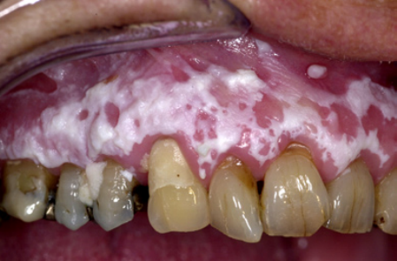
-3.3 Frictional “keratosis”
Another possible reversible white lesion is the frictional
lesion caused by mechanical irritation, e.g. vigorously
brushing of the teeth (Fig. 2). This lesion is sometimes
Fig. 2. Leukoplakia (or "frictional keratosis"?) in a 57-year-old wom-
an.
referred to as “frictional keratosis”. The term “lesion” is
preferred because “keratosis” is actually a histopatho-
logical term. There may be some overlap with the pre-
viously discussed alveolar ridge keratosis (see ad 3.1).
The suggestion to remove this lesion- particularly when
located on the buccal attached gingiva- from the cat-
egory of leukoplakia seems rather questionable (9). A
nal diagnosis of frictional lesion can only be applied to
cases where the lesion has disappeared after elimination
of the possible mechanical cause- provided that there
are no symptoms that would require to immediately bi-
opsy the lesion- , within a somewhat arbitrarily chosen
period of no more than 4-8 weeks. In other words, a de-
nitive diagnosis of frictional lesion can only be made
in retrospect.
-3.4 Hairy leukoplakia
The term hairy leukoplakia is a misnomer- but well ac-
cepted in the literature- for various reasons, being 1) it
is a well described entity, particularly by the immuno-
histochemical demonstration of EBV DNA in the koilo-
cytic epithelial cells of a biopsy specimen, 2) it is not a
potentially malignant disorder, and 3) the clinical aspect
is not always “hairy”. Admittedly, it is difcult to come
up with a better term (10).
-3.5 Restoration associated lesion
A somewhat similar approach as discussed above with
regard to frictional lesions is valid in case of a provision-
al diagnosis of a “contact lesion”, supposedly caused by
direct prolonged contact by large amalgam restorations,
particularly in case of a buccal or lingual extension. A
nal diagnosis of amalgam associated lesion should
only be applied when the lesion has disappeared after
replacement or removal of the amalgam restoration-
provided that there are no symptoms that would require
to immediately biopsy the lesion- , within a somewhat
arbitrarily chosen period of no more than 4-8 weeks.
Therefore, a denitive diagnosis of amalgam associated
lesion can only be made in retrospect.
-3.6 Smokers’ lesion versus tobacco associated leuko-
plakia
It is well known that leukoplakia in patients with to-
bacco habits might be reversible if patients give up their
smoking habits (11). In the absence of symptoms, be-
ing a strong indication for an immediate biopsy in order
to exclude the presence of severe epithelial dysplasia
or even squamous cell carcinoma, the patient should
be advised to give up the tobacco habit. If successful
and if the white lesion regresses within a somewhat ar-
bitrarily chosen period of no more than 4-8 weeks the
provisional clinical diagnosis of such lesion should, in
retrospect, be changed into “smokers’ lesion”. When the
patient is not able or willing to give up the tobacco habit
and in case of persistence of the leukoplakia, the term
“tobacco-associated leukoplakia” can be applied, irre-
spective of the relevance of such designation.
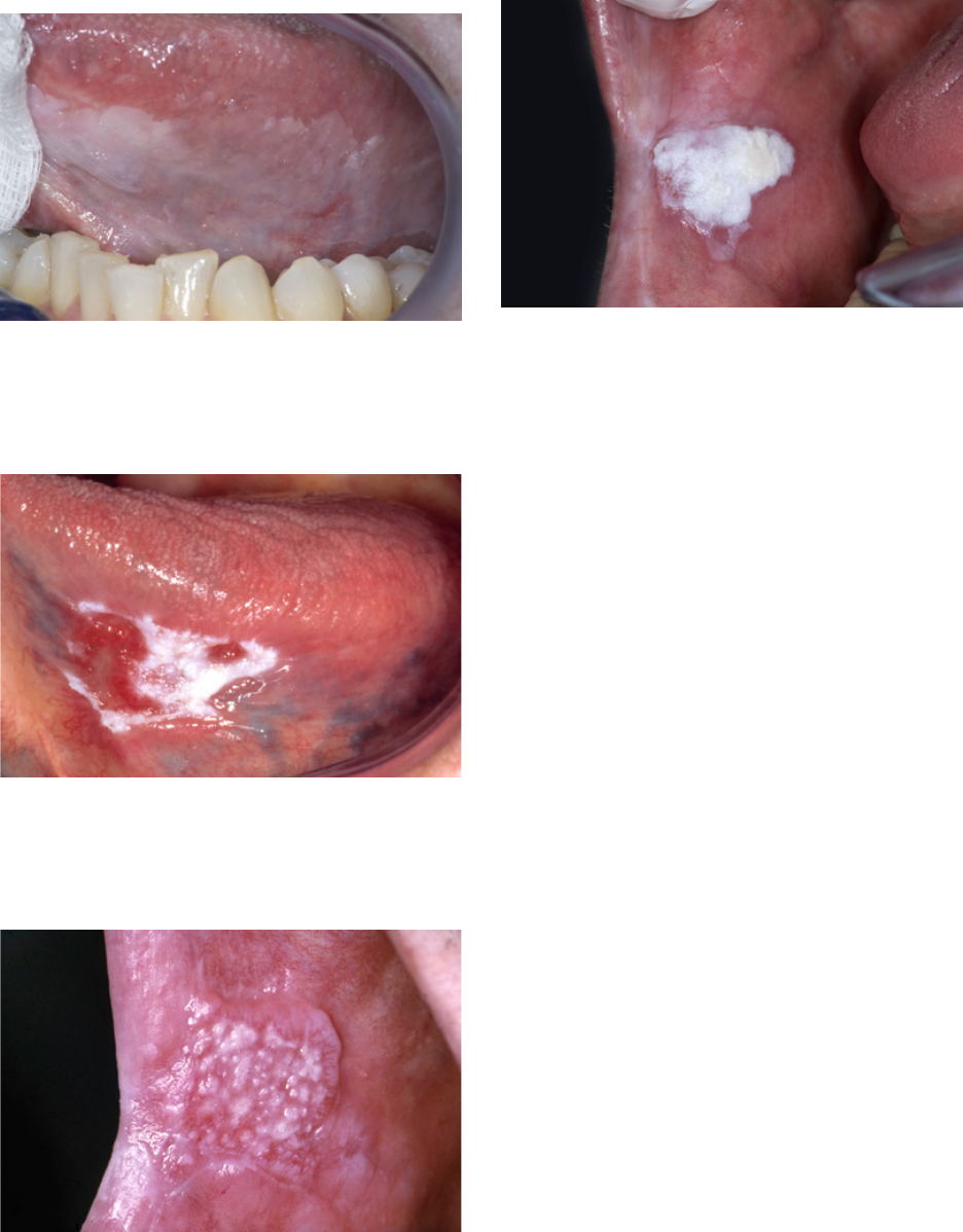
Clinical classication of leukoplakia with em-
phasis on (proliferative) verrucous leukoplakia.
4 1. Traditionally, two major clinical types of leukopla-
kia are recognized, being the homogeneous and the non-
homogeneous type respectively. The signicance of this
classication is the assumption that there is a correla-
tion between the clinical type and the risk of malignant
transformation, the non-homogeneous type carrying a
higher risk. In some studies there is such correlation
while in other studies there is not.
The homogeneous type is characterized by a thin, at
and homogeneous whitish appearance (Fig. 3). The
non-homogeneous type is subdived in a variety of sub-
types, such as speckled or erythematous (white and red
changes), also referred to as erythroleukoplakia (Fig.
4), nodular (Fig. 5) and verrucous (Fig. 6). Particularly
the verrucous type is probably quite often misdiag-
nosed by clinicians because of its homogeneous white
appearance and its often homogeneous (verrucous) tex-
ture. There are actually no strict criteria how to make
a distinction clinically between verrucous leukoplakia
and verrucous carcinoma.
Another confusing type is the proliferative verrucous
leukoplakia (PVL), as being introduced in the literature
by Hansen et al. (12). In the original publication PVL
has been characterized as a slow-growing, persistent,
and irreversible lesion, resistant to all forms of therapy
as recurrence is the rule. PVL may start as a simple
keratosis at one end to invasive carcinoma at the other.
Med Oral Patol Oral Cir Bucal. 2015 Nov 1;20 (6):e685-92. Oral leukoplakia
e690
In fact, a diagnosis of PVL can only be made in retro-
spect. In several of such cases the initial lesion may just
be a solitary homogeneous or non-homogeneous leuko-
plakia (13). Unfortunately, in many scientic reports on
PVL just multifocality and involvement of the gingiva
seem to have been used as diagnostic criteria without
paying attention to the history of the disease.
Some histopathological areas of confusion
-5.1 The assessment of epithelial dysplasia
It is well recognized that the assessment of the presence
and degree of epithelial dysplasia carries a substantial
degree subjectivity, reected in a distinct intra- and in-
terobserver variation (14-16). Unfortunately, in spite of
numerous attempts, as being suggested in the literature,
there is no international consensus on this issue.
Probably some pathologists will deny a diagnosis of
leukoplakia in the absence of epithelial dysplasia, what
is not in accordance with the recommendations from
the “dental” literature.
-5.2 Lichenoid dysplasia
In 1985 the supposedly distinct entity of lichenoid dys-
plasia has been introduced in the literature (17). The
use of of this term is discouraged since it, erroneously,
may suggest dysplastic changes occurring in oral li-
chen planus. Probably a number of the reported cases
of malignant transformation of lichen planus are caused
by forementioned confusing terminology, while cases
of leukoplakia with a lichenoid appearance histopatho-
logically, mainly consisting of a subepithelial bandlike
inltrate, may have erroneously been reclassied as li-
chen planus.
-5.3 Verrucous hyperplasia versus verrucous carcino-
ma
Fig. 3. Homogeneous (at and thin) leukoplakia in a 53-year-old
man.
Fig. 4. Non-homogeneous (white and red changes, also referred to as
erythroleukoplakia) in an 88-year-old woman.
Fig. 5. Non-homogeneous, nodular, leukoplakia in a 61-year-old
man.
Fig. 6. Non-homogeneous, verrucous leukoplakia. In spite of a ho-
mogeneous white appearance and a homogeneous verrucous texture,
this lesion should not be called homogeneous leukoplakia
Med Oral Patol Oral Cir Bucal. 2015 Nov 1;20 (6):e685-92. Oral leukoplakia
e691
Several papers have been published about the his-
topathological difference between verrucous hyper-
plasia and verrucous carcinoma, still leaving room for
discussion (18,19). In daily practice it is difcult, if not
impossible, to make this distinction in a reproducible
way. Besides, one may question the clinical relevance of
the distinction between these two entities since for both
lesions (surgical) removal is recommended. A pitfall is
that some pathologists may describe these epithelial
changes as being benign, while the behavior of such le-
sions actually is unpredictable.
Discussion and Conclusions
It is well appreciated that a number of aspects of the
presently discussed denition and terminology may not
be equally valid in all parts of the world. A classica-
tion of potentially malignant disorders has been pro-
posed in 2011 from India (20). Apparently, this classi-
cation is not limited to leukoplakia, but also includes
entities such as lichen planus, oral submucous brosis,
nutritional deciencies and some inherited cancer syn-
dromes.
The recommendation is made to modify the present
2005 WHO denition of oral leukoplakia, amongst oth-
ers by adding explicitly the requirement of histopatho-
logic examination in order to obtain a denitive clin-
icopathological diagnosis. As a result, the following
denition is proposed: “A predominantly white patch
or plaque that cannot be characterized clinically or
pathologically as any other disorder; oral leukoplakia
carries an increased risk of cancer development either
in the area of the leukoplakia or elsewhere in the oral
cavity or the head-and-neck region”.
Furthermore, the use of strict diagnostic criteria is rec-
ommended for predominantly white lesions or diseases
for which a possible causative factor has been identied,
e.g. smokers’ lesion, frictional lesion and dental resto-
ration associated lesion. An observation of 4-8 weeks
after removal of the suggested cause seems a practical
one and seems also safe practice, particularly in case of
an asymptomatic leukoplakic disorder. In this respect
one should realize that at the rst visit of a patient with
oral leukoplakia a squamous cell carcinoma may be
present already and one would not run the risk of ob-
serving such event for a period of more than 4-8 weeks.
Even such period is already a long one in case of a sq-
uamous cell carcinoma, a carcinoma in situ or severe
epithelial dysplasia. However, it should be emphasized,
that the presence of such changes is nearly always as-
sociated with symptoms. Therefore, in the presence of
symptoms a biopsy is strongly recommended before
elimination of possibly causative factors and observa-
tion of the result of such elimination.
As is true for almost all pathologies proper communica-
tion between clinicians and pathologists is important,
particularly in the eld of oral potentially malignant
disorders. For instance, some pathologists will deny
a diagnosis of leukoplakia in the absence of epithelial
dysplasia. Also the use of the term “lichenoid dysplasia”
may be the subject of confusion between pathologists
and clinicians.
References
1. Kramer IR, Lucas RB, Pindborg JJ, Sobin LH. Denition of leu-
koplakia and related lesions: an aid to studies on oral precancer. Oral
Surg Oral Med Oral Pathol. 1978;46:518-39.
2. Pindborg JJ, Reichart PA, Smith CJ, van der Waal I. World Health
Organization International Histological Classication of Tumours.
Histological Typing of Cancer and Precancer of the Oral Mucosa.
Second Edition ed. Berlin, Heidelberg, New York: Springer-Verlag.
1997.P. 1-85.
3. van der Waal I, Axéll T. Oral leukoplakia: a proposal for uniform
reporting. Oral Oncol. 2002;38:521-6.
4. Brouns EREA, Baart JA, Bloemena E, Karagozoglu KH, van der
Waal I. The relevance of uni-form reporting in oral leukoplakia: De-
nition, certainty factor and staging based on experience with 275
patients. Med Oral Patol Oral Cir Bucal. 2013;18:e19-26.
5. Warnakulasuriya S, Johnson NW, van der Waal I. Nomenclature
and classication of potentially malignant disorders of the oral mu-
cosa. J Oral Pathol Med. 2007;36:575-80.
6. Pindborg JJ, Jolst O, Renstrup G, Roed-Petersen B. Studies in oral
leukoplakia: a preliminary report on the period pervalence of malig-
nant transformation in leukoplakia based on a follow-up study of 248
patients. J Am Dent Assoc. 1968;76:767-71.
7. Chi AC, Lambert PR, Pan Y, Li R, Vo DT, Edwards E et al. Is
alveolar ridge keratosis a true leukoplakia? A clinicopathologic com-
parison of 2,153 lesions. J Am Dent Assoc. 2007;138:641-51.
8. Natarajan E, Woo SB. Benign alveolar ridge keratosis (oral lichen
simplex chronicus): A distinct clinicopathologic entity. J Am Acad
Dermatol. 2008;58:151-7.
9. Mignogna MD, Fortuna G, Leuci S, Adamo D, Siano M, Makary
C et al. Frictional keratoses on the facial attached gingiva are rare
clinical ndings and do not belong to the category of leu-koplakia. J
Oral Maxillofac Surg. 2011;69:1367-74.
10. Van der Waal I. Greenspan lesion is a better term than oral
“hairy” leukoplakia. J Oral Pathol Med. 1996;25:144.
11. Warnakulasuriya S, Dietrich T, Bornstein MM, Casals PE, Pres-
haw PM, Walter C et al. Oral health risks of tobacco use and effects
of cessation. Int Dent J. 2010;60:7-30.
12. Hansen LS, Olson JA, Silverman S. Proliferative verrucous leu-
koplakia. A long-term study of thirty patients. Oral Surg Oral Med
Oral Pathol. 1985;60:285-98.
13. Van der Waal I, Reichart PA. Oral proliferative verrucous leuko-
plakia revisited. Oral Oncol. 2008;44:719-21.
14. Warnakulasuriya S, Reibel J, Bouquot J, Dabelsteen E. Oral
epithelial dysplasia classication systems: predictive value, uti-
lity, weaknesses and scope for improvement. J Oral Pathol Med.
2008;37:127-33.
15. Abbey LM, Kaugars GE, Gunsolley JC, Burns JC, Page DG,
Svirsky JA, et al. Intraexaminer and interexaminer reliability in the
diagnosis of oral epithelial dysplasia. Oral Surg Oral Med Oral Pa-
thol Oral Radiol Endod. 1995;80:188-91.
16. Kujan O, Oliver RJ, Khattab A, Roberts SA, Thakker N, Sloan
P. Evaluation of a new binary system of grading oral epithelial
dysplasia for prediction of malignant transformation. Oral Oncol
2006;42:987-93.
17. Krutchkoff DJ, Eisenberg E. Lichenoid dysplasia: a distinct histo-
pathologic entity. Oral Surg Oral Med Oral Pathol. 1985;60:308-15.
18. Shear M, Pindborg JJ. Verrucous hyperplasia of the oral mucosa.
Cancer. 1980;46:1855-62.
19. Murrah VA, Batsakis JG. Proliferative verrucous leukoplakia and
verrucous hyperplasia. Ann Otol Rhinol Laryngol. 1994;103:660-3.
Med Oral Patol Oral Cir Bucal. 2015 Nov 1;20 (6):e685-92. Oral leukoplakia
e692
20. Sarode SC, Sarode GS, Karmarkar S, Tupkari JV. A new classi-
cation for potentially malignant disorders of the oral cavity. Oral
Oncol. 2011;47:920-21.
Conict of interest
None declared
